Introduction
This article analyzes the results of using a keyhole approach to treat 30 cases (diameter ≤3 cm) of intracranial minimally invasive lesions (navigation group) under the guidance of Heal Force Excelim-04 neurosurgical system at the First Affiliated Hospital of Guiyang Medical College. At the same time, 25 cases (microsurgery group) with simple microsurgery were used for multiple index control. It was concluded that keyhole surgery is superior to traditional microsurgery in the treatment of intracranial microlesions with the aid of a neuronavigation system.
Clinical Results
(30 patients in the navigation group and 25 patients in the microsurgery group)
The navigation average registration error (2.10 ± 0.52) mm. Compared with simple microsurgery, there was a statistically significant difference in bone diameter, scalp incision length, postoperative complications, and days of hospital stay in the navigation group (all P<0.05). Followed up for 1 year. In the navigation group and microsurgery group, the condition improved or no change occurred in 24 cases and 18 cases respectively, and the aggravation was 5 cases and 7 cases respectively. One case died in the navigation group.
Patient Data
Clinical data and inclusion criteria Case inclusion criteria: 1 Single intracranial lesion. 2 lesion diameter ≤ 3 cm.
Using navigation guided keyhole surgery (navigation group) in 30 cases::Including 19 males and 11 females; aged 4 to 72 years, mean 42.4 years old. CT or MRI showed a lesion diameter of 0.6 to 3.0 cm, with an average of 2.1 cm. Preoperative symptoms: 11 cases of headache, 3 cases of limb weakness, 4 cases of epilepsy, 1 case of language impairment, 3 cases of vision loss.
Using simple microsurgery (microsurgery group) in 25 cases:Including 14 males and 11 females; aged 18 to 70 years, mean 47.9 years old. CT or MRI showed a diameter of 1.0 to 3.0 cm with an average of 2.4 cm. Preoperative symptoms: 12 cases of headache, 1 case of facial facial pain, 2 cases of hearing loss on one side, 3 cases of weakness on one side, and 1 case of vision loss.
Research Methods
Clinical Method:Using Excelim-04 surgical navigation system. The navigation team shaved their heads 1 day before surgery. The scalp markings (Marker) were affixed to the imaging system and imported into the navigation system. The registration error was generally <3.0 mm. The microsurgery group did not use navigation-assisted positioning, and the rest of the operations were in the navigation group.
Statistical Method:Use SPSS16.0 statistical software. The measurement data is expressed as x ± s, which is in accordance with the normal distribution using the t-test. The non-parametric test (Mann-whitney U test) is not applicable to the normal distribution. The chi-square test is used to compare the two sample rates. The difference was statistically significant at P<0.05.
Result Analysis
【Navigation Results】
The navigation average registration error (2.10 ± 0.52) mm, intraoperative navigation and positioning accuracy, all lesions are directly to the lesion.

【Surgical Efficacy】
Compared with the microsurgery group, the diameter of the bone window, scalp incision length, postoperative complications, and length of hospital stay in the navigation group were statistically significant (all P<0.05), but there was no significant difference in the maximum diameter of the lesion and the operation time (both P>0.05). Postoperative patients can take care of themselves.
Navigation Group:Four patients developed complications, including 1 case of motor language disorder, 2 cases of limb paralysis, and 1 case of limb dysfunction.
Microsurgery Group:Complications occurred in 9 cases, including 3 cases of transient urinary collapse, 4 cases of limb paralysis, and 2 cases of scalp wound infection.

【Follow-up Results】
After 1 year of follow-up, the navigation group improved or had no change in 24 cases, aggravated in 5 cases, and died in 1 case. Microsurgery group improved or no change in 18 cases, 7 cases of increased symptoms.
Postoperative Analysis
At present, there is no uniform standard for the size of intracranial microlesions. The maximum diameter of the lesions reported in the literature is about 3cm. Considering the surgical operation space, the diameter of the lesion treated by keyhole surgery is better than 3 cm.
The advantages of using neuronal navigation to use the keyhole approach to treat intracranial microlesion are as follows:
①Targeting
In the operation of keyhole surgery for intracranial micro-lesions, accurate positioning is the key to successful operation. Targeted positioning can solve the embarrassing situation that the lesion can not be found after the routine operation of the craniotomy, and it is forced to increase the length of the scalp incision and the size of the bone window due to the difficulty in positioning.
②Effectively avoid important blood vessels, nerves, determine tumor boundaries
Microscopic low-grade astrocytomas, cavernous hemangioma lesions, and normal brain tissue are difficult to distinguish under the naked eye. During surgery, multiple sites are required to take frozen biopsies to define the extent of the tumor. The neuronavigation system can accurately determine the extent of the tumor and guide surgical resection. In the resection of a large part of the recurrence of large pituitary tumors, the normal anatomy is often unclear, neural navigation can effectively avoid important structures, determine the tumor boundary.
Problems with neuronavigation:
Image drift
This is a persistent problem with neuronavigation. However, when using neuronavigation aids for keyhole surgery, the impact of structural image drift is limited due to the small bone windows and low release of cerebrospinal fluid. The surgeon should pay more attention to the presence of systemic image drift. If imaging drift is found, for cystic lesions or vascular lesions, intraoperative ultrasound can be used to detect the lesion and find the boundary between the diseased tissue and normal brain tissue; the complex structure around the lesion, such as lesions in the functional area and neighboring or wrapping important fiber bundles At the time, image fusion can be performed using functional magnetic resonance (fMRI) and diffuse tensor fiber tomography (DTT) to maximize brain protection under direct vision.
Summary:
Neural navigation assisted keyhole surgery has broad application prospects in the field of minimally invasive neurosurgery. However, it should be pointed out that the keyhole surgery is not limited to only one small bone hole, but according to the specific conditions of the patient, design the minimal surgical incision that is most suitable for the resection of the lesion. In addition, neuronavigation as an auxiliary device, although it has the advantages of high accuracy and can reduce the difficulty of surgery, but can not replace the surgeon's experience and basic operating techniques, the surgeon's precise operation is the determinant of surgery.
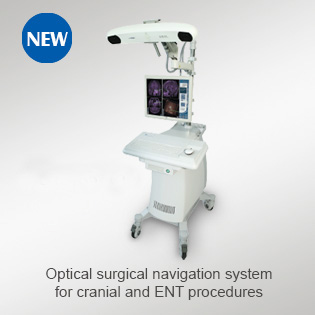
Heal Force Excelim-04 Neurosurgical Navigation System
Rich fusion
Compatible with DTI/PET/DSA/CTA/PCA/MRA/MRV/
T1 MRI/fMRI/Bold MRI/MRS Image fusion and processing
﹀
Clear reconstruction
Reconstructed 2D images and 3D models with clear details
Supports Fusion Modeling of Multiple Medical Image Sequences
﹀
Easy Planning
Can simulate multiple surgical approaches
Tomographic image length measurement
Automatic measurement of lesions
﹀
Accurate Navigation
Positioning error is less than 1mm
Multiple ways to correct brain drift